Lab-grown corneal epithelial cells can potentially be used for restoring vision
Researchers at the Hyderabad-based LV Prasad Eye Institute (LVPEI) have successfully grown miniature eye-like organs that closely resemble the developing eyes of an early-stage embryo. The miniature eyes were produced using induced pluripotent stem (iPS) cells. The iPS cells are produced by genetically manipulating human skin cells to produce embryonic-like stem cells that are capable of forming any cell types of the body.
Small portions of the corneal tissue were separated from the miniature eyes and used for growing corneal epithelial cell sheets in the lab. Such tissue-engineered cell sheets can potentially be used for restoring vision in patients whose limbus region of the cornea is damaged in both the eyes. The limbus region of the cornea contains stem cells, and chemical or thermal damage to this region affects corneal regeneration and results in vision loss.
Stem cells present in the limbus region of a healthy eye have been used for restoring vision when only one eye is damaged. But when the damage is present in both eyes, the only way to restore vision is by using the healthy limbus taken from a related or unrelated donor. Patients have to be on immunosuppressants lifelong when limbus is transplanted from donors. However, immunosuppressants are not required when corneal cells grown using the patient’s own skin cells are used for restoring vision.
Growing eye-like organs
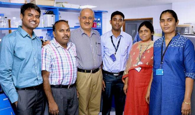
A team led by Dr. Indumathi Mariappan was able to grow complex eye-like organs in the lab by allowing the cells to organise themselves in three dimensions. The miniature eye closely resembles the developing eyes of an early-stage embryo. The eye-like structure consists of miniature forms of retina, cornea and eyelid. The results were published in the journal Development.
“It took about four–six weeks for the eye-like structure to form from iPS cells. We then removed the cornea-like structure for further study,” says Dr. Mariappan from the Centre for Ocular Regeneration at the LV Prasad Eye Institute and the corresponding author of the paper.
The cornea has three layers — epithelium (outer layer), stroma (middle layer) and endothelium (inner layer). “All the three layers of the cornea were observed, indicating that the mini-cornea had developed correctly,” she says. “The cornea initially forms as a simple bubble-like structure which is very delicate to handle. It later matures to form a thick cornea-like structure over a period of 10-15 weeks.”
The corneal epithelial sheets that would be used for treating the damaged eyes were then grown in the lab using small pieces of the mini-cornea containing the epithelium and a portion of the stroma. The stem cells present in the tissue pieces proliferated and gave rise to a uniform sheet of epithelium of about 2.5 cm by 2.5 cm size.
Animal trials
The team is currently focusing on testing the usefulness of the corneal cells grown from iPS cells in restoring vision in animal models (rats). “We will soon be starting the animal experiments,” she says. Trials on human subjects will be considered if the animal experiments turn out to be safe and effective in restoring vision.
In treatment
In parallel, the researchers are also working on producing mini-retinal tissue and actively exploring iPS cell-derived retinal tissues for treating several retinal diseases such as age-related macular degeneration (AMD), retinitis pigmentosa and certain forms of congenital blindness seen in children and young adults.
Already, retinal cells grown using human embryonic stem cells and iPS cells are being tested in clincal trials in a few countries to treat retinal diseases.
source: http://www.thehindu.com / Home> Sci-Tech> Health / by R. Prasad / June 17th, 2017